Il Prof Ungar attraverso il suo slide show ci fornisce un excursus sul mondo della sincope e delle perdite di coscienza aiutando il professionista dell’ emergenza urgenza a muoversi appropriatamente in questo argomento, tema che molte volte genera dei caratteristici ” TOUR DELLA SINCOPE”
La Drssa Bagatti, anestesista rianimatore , ci conduce per mano all’interno del mondeo della DONAZIONE .
LA donazione degli organi negli ultimi annoìi ha assunto un ruolo strategico nel processo di cura di particolari patologie – Aver costituito una eccellenza sanitaria porta ad outcome favorevoli dei pazienti.
La Drssa Bagatti rappresenta un punto di riferimento di quest delicatissimo tema ed ha portato la propri azienda sanitaria ( USL PRATO poi Toscana Centro) a livelli di eccellenza , anche grazie alla collaborazione ed al contributo dei Pronto Soccorso
A 57 year-old-patient is brought in Emergency Department by ambulance complaining of fever, swelling and erythema of the left leg .
Medical History: not relevant.
Vital Signs: BP 130/80 mmHg, HR 90 bpm, SpO2 96% room air, RR 22/min, GCS 15, T 38°C.
Arterial Blood Gas analysis:
ECG. 1
ECG ANSWER and INTERPRETATION
Rate:
85 min
Rhythm:
Regular P waves
Axis:
-75°
Intervals:
PR – normal (<200ms) and regular
QRS – Wide (>120ms); rSR’ morphology V1-V3: RBBB.
QT – 360ms (QTc Bazett 430ms)
Waves and Segments:
P waves: left atrial abnormality (negative component>positive component)
Q waves DII-DIII-aVF: >0.04ms and Q>R
S wave in DI
ST depression V1-3 and DI-aVL
T waves: negative V1-V3, DIII, aVR
SIQIIITIII (McGinn-White): S wave in DI, Q waves DIII, negative T waves in DIII
DIFFERENTIAL DIAGNOSIS
The ECG changes described may be seen with any cause of acute or chronic disease that causes right ventricular strain:
Acute
Chronic
Severe pneumonia Exacerbation of COPD /asthma Pneumothorax Recent pneumonectomy Upper airway obstruction Pulmonary Embolism
Chronic obstructive pulmonary disease Recurrent small PEs Cystic fibrosis Interstitial lung disease Severe kyphoscoliosis Obstructive sleep apnoea Pulmonary Ipertension
OUTCOME
There were no focal changes in lung parenchyma on chest radiographs.
The patient was allergic to iodinated contrast media (anaphylactic shock): A pulmonary scintigraphy was then performed in the suspicion of pulmonary embolism.
IMG. 1-4: Pulmonary scintigraphy
Ventilation images document areas of impaired ventilation in the posterior basal area of the left lung; Perfusion images document perfusion deficit in the posterior basal segment.
Conclusion: compatible with pulmonary embolism.
The patient was treated with Apixaban and discharged after a few days.
TRICKS
ECG changes associated with acute pulmonary embolism are nonspecific and may be seen in other condition [1-5]. The ECG in Pulmonary Embolism (PE) lacks sensitivity and specificity: it is neither sensitive nor specific enough to diagnose or exclude PE.
ECG changes in PE are related to dilation of the right atrium and right ventricle, right ventricular ischaemia, heart rotation and position shift, increased sympathetic tone due to pain, anxiety and hypoxia.
The most common findings are sinus tachycardia (44%) and nonspecific ST-segment and T-wave changes (50 %). The most specific finding is simultaneous T wave inversions in the inferior (II, III, aVF) and right precordial leads (V1-4), representing high pulmonary artery pressures; T wave inversion in lead V1 plus lead III was only seen in 1% of ACS patients versus 88% of patients with PE.
Other ECG changes in pulmonary embolism include:
Complete or incomplete RBBB (18%)
Right ventricular strain pattern – T wave V1-4 ± II, III, aVF
Right axis deviation – (16%)
Dominant R wave in V1 – representing acute right ventricular dilatation.
Right atrial enlargement – P wave in lead II > 2.5 mm (9%)
SIQIIITIII –neither sensitive nor specific. Abnormalities historically considered to be suggestive of PE (S1Q3T3 pattern, right ventricular strain, new incomplete right bundle branch block) are uncommon (less than 10 percent) [6-7].
Clockwise rotation
Atrial tachyarrhythmias – AF, flutter, atrial tachycardia (8%)
Normal ECG is present in 9-26% of cases.
ECG abnormalities that are associated with a poor prognosis [1,2,4]:
●Atrial arrhythmias
● Right heart strain + RBBB
●Bradycardia (<50 beats per minute) or tachycardia (>100 beats per minute)
●Complete right bundle branch block
●Inferior Q-waves (leads II, III, and aVF)
●Anterior ST-segment changes and T-wave inversion (V1-V3)
●SIQIIITIII
AUTHORS
Dr. Lorenzo Pelagatti
Emergency Department – Nuovo Ospedale di Prato S. Stefano – Prato, Tuscany, Italy
High Dependency Unit – AOU Careggi – Florence, Tuscany, Italy
Dr. Franco Lai
Emergency Department – Nuovo Ospedale di Prato S. Stefano – Prato, Tuscany, Italy
REFERENCES
Geibel A, Zehender M, Kasper W, Olschewski M, Klima C, Konstantinides SV. Prognostic value of the ECG on admission in patients with acute major pulmonary embolism. Eur Respir J. 2005 May;25(5):843-8. doi: 10.1183/09031936.05.00119704. PMID: 15863641.
Ferrari E, Imbert A, Chevalier T, Mihoubi A, Morand P, Baudouy M. The ECG in pulmonary embolism. Predictive value of negative T waves in precordial leads–80 case reports. Chest. 1997 Mar;111(3):537-43. doi: 10.1378/chest.111.3.537. PMID: 9118684.
Rodger M, Makropoulos D, Turek M, Quevillon J, Raymond F, Rasuli P, Wells PS. Diagnostic value of the electrocardiogram in suspected pulmonary embolism. Am J Cardiol. 2000 Oct 1;86(7):807-9, A10. doi: 10.1016/s0002-9149(00)01090-0. PMID: 11018210.
Shopp JD, Stewart LK, Emmett TW, Kline JA. Findings From 12-lead Electrocardiography That Predict Circulatory Shock From Pulmonary Embolism: Systematic Review and Meta-analysis. Acad Emerg Med. 2015 Oct;22(10):1127-37. doi: 10.1111/acem.12769. Epub 2015 Sep 22. PMID: 26394330; PMCID: PMC5306533.
Co I, Eilbert W, Chiganos T. New Electrocardiographic Changes in Patients Diagnosed with Pulmonary Embolism. J Emerg Med. 2017 Mar;52(3):280-285. doi: 10.1016/j.jemermed.2016.09.009. Epub 2016 Oct 11. PMID: 27742402.
Panos RJ, Barish RA, Whye DW Jr, Groleau G. The electrocardiographic manifestations of pulmonary embolism. J Emerg Med. 1988 Jul-Aug;6(4):301-7. doi: 10.1016/0736-4679(88)90367-8. PMID: 3225435.
Thames MD, Alpert JS, Dalen JE. Syncope in patients with pulmonary embolism. JAMA. 1977 Dec 5;238(23):2509-11. PMID: 578884
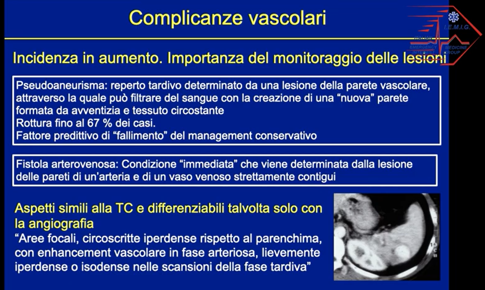
Una lezione sui traumi addominali chiusi a cura della Dott.ssa Stefania Salti e Dott. Andrea Bulli – UO Radiologia, Nuovo Ospedale di Prato Santo Stefano.
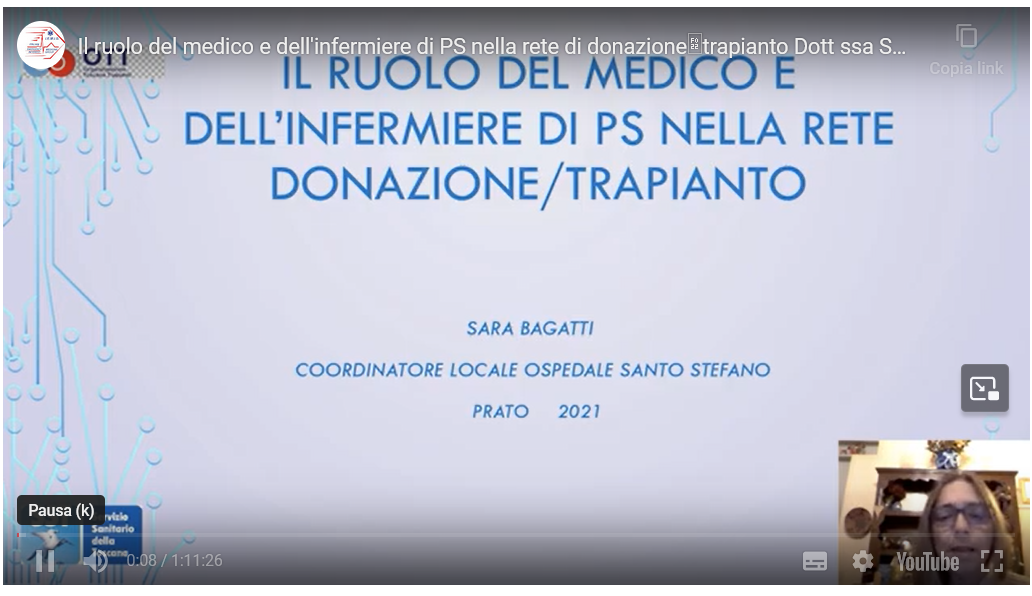
A cura della Dott.ssa Sara Bagatti, Coordinatrice locale alla donazione di organie tessuti del Nuovo Ospedale Santo Stefano di Prato e dell’Azienda Ospedaliero Universitaria Careggi, nonchè coordinatrice dell’Area Vasta Toscana Centro, un interessante panoramica e disamina sul ruolo medico ed infermieristico nella rete di donazione e trapianto in Dipartimento di Emergenza- Urgenza.
Grazie ad AMI ( associazione ONLUS Materno Infantile . Ospedale Santo Stefano – PRATO) – IEMIG ha partecipato alla Giornata della FESTA DELLA MAMMA” , manifestazione tenutasi nei nuovi giardini dell’ Ospedale di Prato. Attraverso il coinvolgimento del magnifico mondo del volontariato pratese , con la partecipazione di MISERICORDIA PUBBLICA ASSISTENZA ; CROCE D’ ORO e CROCE ROSSA, si sono tenute dimostrazioni sulle principali manovre di soccorso e di life saving.
– Regular – Rate ~140-280 bpm – QRS complexes have a morphology that is usual for the subject under examination – Narrow QRS complexes (< 120 ms) unless there is co-existing bundle branch block, accessory pathway, or rate-related aberrant conduction – P waves if visible exhibit retrograde conduction with P-wave inversion. – P waves if visible exhibit retrograde conduction with P-wave inversion in leads II, III, aVF. They may be buried within (more frequently), visible after, or very rarely visible before the QRS complex – The atrial and ventricular rates are the same – ST-segment depression may be observed
AVRT
– Regular – Rate 200-300 bpm – QRS complexes have a morphology that is usual for the subject under examination – Narrow QRS complexes (< 120 ms) unless there is co-existing bundle branch block, accessory pathway, or rate-related aberrant conduction – P waves if visible exhibit retrograde conduction with P-wave inversion. – Retrograde P waves are usually visible, with a long RP interval (P-QRS > QRS-P)QRS < 120ms unless pre-existing bundle branch block, or rate-related aberrant conduction – The atrial and ventricular rates are the same – ST-segment depression may be observed
Atrial Flutter
– Narrow complex tachycardia – Regular atrial activity at ~250-300 bpm (more frequently ~300 bpm) – “Saw-tooth” pattern of P waves (typically in II, III, aVF, V1) – Upright flutter waves in V1 that may resemble P waves – Ventricular rate depends on AV conduction ratio; QRS rate is a fraction of the atrial rate (1/2 = 150 bpm; 1/3 = 100 bpm: 1/4 = 75 bpm)
OUTCOME
Flutter waves are not clearly seen (flutter with a 1:1 block?); Alternatively, this may just be rapid SVT (AVNRT/AVRT).
Given doubt regarding the rhythm, and a fixed heart rate of 220 bpm that is often seen in atrial flutter, we chose to administer adenosine to assist in differentiating/ treating the cause.
6 mg of adenosine was administered without benefit.
Then 12 mg of adenosine was administered.
ECG 2.
Rhythm strip reveals flutter waves. The patient does not cardiovert to sinus rhythm following adenosine administration; instead, the degree of AV block is increased, revealing sawtooth flutter waves.
Verapamil 5 mg 1 fl + Normal Saline 250 ml was administered, with good rate control
Per laureati nel corso di laurea in medicina e chirurgia, corso di laurea in infermieristica, e neospecialisti della scuola di specializzazione in medicina di emergenza-urgenza.
Le emergenze gastroenterologiche in PS rappresentano una importante “fetta” di casistica che impegna risorse umane e tecnologiche; molte volte assistiamo a presentazioni cliniche che intercettano le reti tempo dipendenti .Il Dr Marchi in questa presentazione traccia un excursus sulle principali emergenze gastroenterologiche
L embolia polmonare in PRONTO SOCCORSO è una delle diagnosi più sfidanti, presentandosi molte volte con pattern sintomatologici e semeiotici subdoli. La Drssa Ottaviani presenta un excursus sul processo diagnostico
Utilizzando il sito, accetti l'utilizzo dei cookie da parte nostra. maggiori informazioni
Questo sito utilizza i cookie per fornire la migliore esperienza di navigazione possibile. Continuando a utilizzare questo sito senza modificare le impostazioni dei cookie o cliccando su "Accetta" permetti il loro utilizzo.